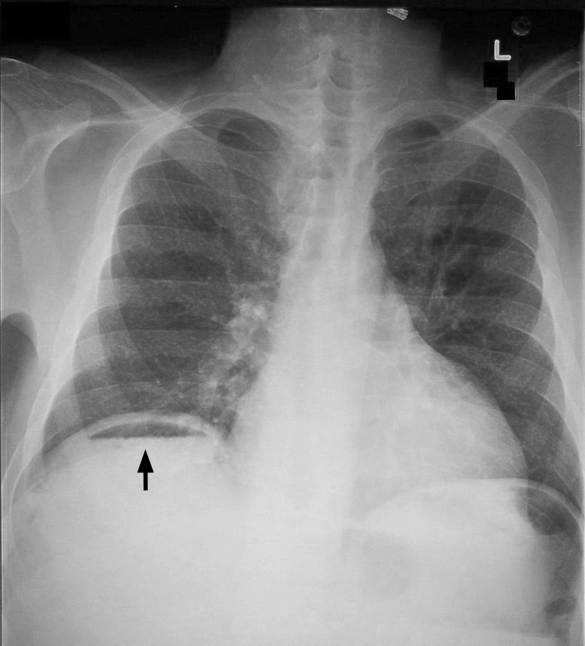
Examples of plain radiography in adults example 1 and example 2 of a male adult’s chest radiograph Chest X-Ray Normal Findings Clear the costophrenic angles and lung fields.
We try our best to provide photographs with the best quality or high resolution possible. I purchased a few of papers here, and they were all excellent. TWO VIEWS OF A CHEST X-RAY. This makes sense and applies the inductive approach.
How to write normal chest x ray report.

Chest X Ray Interpretation A Structured Approach Radiology Osce How To Write Job Progress Report Writing Example On Earthquake
The report serves as a legal document and shows the radiologist’s attitude, perception, and skill. The left lower hemithorax’s base has a faint, rounded density that is most likely a nipple shadow. Teaching Lessons Using Chest X-Rays.
The radiologist and the referring doctor communicate mostly through the radiological report. Learn more about the stunning advantages that demonstrate our unwavering commitment to our clients. Right-sided preponderance causes the cardiac shadow to be bigger.
These zones do not correspond to lung lobes, for example. There is no proof of a consolidation focus area. A brief note to prevent confusion
Each lung should be divided into three zones, each taking up one-third of the lung’s height, when interpreting a chest X-ray. An X-ray of the chest showing the silhouette sign. The report is the written representation of the radiologists’ analysis, recommendations, and interpretation of the radiologic study.
Extrathoracic soft tissue effusions. A decision is reached after weighing the information. No, the patient is not the same 3.
Because it’s free to uncover pertinent material, you can use background sources without paying for them. NB The chest may be expanded due to COPD, then write. Principles of Reading 1.
Examples of typical imaging of the chest and nearby regions are provided in this article, organized by modality. X-rays are seen such that the patient’s right side, as if facing you, is on the left side of the image. Check the location of the hemidiaphragms. Due to the liver, the right is often slightly higher than the left. In cases of tension pneumothorax or aspiration of a foreign body, the contour may flatten unilaterally or bilaterally. For free gas, look underneath the diaphragm.
The most common imaging procedure in the United States is an X-ray of the chest, usually known as a chest radiograph or chest radiograph.
Despite other diagnostic imaging laboratory tests and extra physical examinations, it is almost often the initial imaging study ordered to evaluate for thoracic diseases. Students will gain an understanding of the range of normal markings, the fundamentals of CXR reading, and how patient age and sex affect differentials by reading this series of Normal CXRs. Is the patient in this lateral chest x-ray the same as the patient in the PA film?
April 24th, 2021, report For your benefit, we’ve tried to locate the most effective references about Sample Radiology Report Pdf And How To Write A Chest X Ray Report. If not, then also indicate that in the report.
Only two lobes, but three zones, make up the left lung. Fever, aches, and symptoms similar to the flu Verify if lung marks are present across the lung zones by inspecting them.
We like it because it came from a reliable online source. many items, including pacemakers, catheters, etc. a convenient website with quick service and good papers.
Yes, patient 2 is the same. Topic research, writing, editing, and proofreading are included. formatting How To Write Normal Chest X Ray Report plagiarism scans and subsequent edits.

How To Read Chest X Rays International Emergency Medicine Education Project What Is Subject Knowledge Report Cover Page Template Word Free


